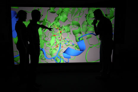
FAYETTEVILLE, Ark. – To understand a protein, it helps to get inside of it, and a University of Arkansas professor has figured out a way to do so.
James F. Hinton, University Professor of chemistry and biochemistry, has worked with Virtalis, an advanced visualization company, to create a computer software program and projection system that lets a person look at larger-than-life, 3-D structures of proteins in virtual reality. This allows scientists to walk inside, through or around the protein of interest for investigating its structure and function.
“Proteins are very complex molecular structures,” said Hinton. Proteins are built from amino acids, molecules that share certain characteristics and have unique side chains. Yeast proteins can have 466 amino acids, while the larger proteins have almost 27,000 amino acids. These amino acids interact to form a particular structure for each protein, and this structure helps to determine the function of the protein.
Since proteins underlie most human diseases, they interest researchers studying the underlying mechanisms of disease. The flu virus, for instance, harbors proteins that cause the illness experienced by humans. The bacterium Staphylococcus aureus produces a toxic protein that causes many of the symptoms experienced by the body. Figuring out how to neutralize these proteins could help treat or prevent disease.
 Virtual reality allows researchers to simulate modifications to proteins and determine possible outcomes.
|
Scientists find that examining protein interactions in two dimensions ranges from tedious to impossible because of the proteins’ size and complexity. Hinton worked with the advanced visualization company Virtalis to develop the ActiveMove Virtual Reality system for PyMOL, a three-dimensional molecular viewing program. The Virtalis system allows researchers to enlarge the protein to room-size and examine it from all sides, including the inside, which can be crucial for understanding the relationship between structure and function.
“Using this system, we can answer many questions about interactions. Why does a toxic protein do what it does? Does the protein form a channel? If it does, what does it look like? And how can we block it?” Hinton said. “This system can act as a guide for what to do next.”
Many proteins, such as a mushroom-shaped toxin from Staphylococcus aureus, form channels to perform their functions and carry out their interactions through binding to other proteins. By virtually exploring the proteins, scientists can determine what kinds of interactions might block the toxic functions of such a protein, or make virtual modifications to the proteins themselves to see if the modifications render them unable to interact and bind to other proteins.
“Thanks to the National Institutes of Health, which has funded the University’s Center for Protein Structure and Function for many years, we have superb instrumentation,” Hinton said. “The immersive Virtual Reality System provides us with another way of enhancing the data we get from those instruments.”
The ActiveMove system includes a 3-D projector with a rear projections screen, coupled with a personal computer, eyewear, head and hand tracking and Virtalis software and support. Funds from the Arkansas Biosciences Institute were used to purchase the Virtalis Virtual Reality System.
Contacts
James F. Hinton, University Professor, chemistry and biochemistry
J. William Fulbright College of Arts and Sciences
479-575-5143,
Melissa Blouin, director of science and research communication
University Relations
479-575-3033,