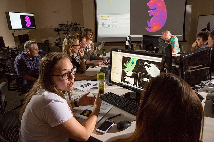
Researchers in the J. William Fulbright College of Arts and Sciences and the College of Engineering recently installed and tested the University of Arkansas' new MicroCT imaging system located in the J.B. Hunt Center of Academic Excellence Building.
Commonly referred to as "CT" — the same sort of scanning used for medical-imaging — computed tomography uses X-ray technology to generate high-resolution 2-D and 3-D representations of an object's internal and external structure. MicroCT produces images that allow researchers to examine materials down to the micro- (less than or equal to 0.1 millimeter) and even nano-scale (less than 0.001 millimeter).
.jpg) Claire Terhune, assistant professor of anthropology. |
MicroCT enables researchers to visualize bones, teeth and archeological artifacts up close and internally, without dismantling or destroying them, and will thus help researchers gain new and exciting information about evolution, human behavior and cognitive function. But these applications only scratch the surface.
MicroCT can also be used for analysis of additive-manufacturing techniques, aerospace technologies, biomedically engineered bone and soft tissue structures, and many other items — basically any object the size of a basketball or smaller and weighing no more than 110 pounds.
Managed by the Center for Advanced Spatial Technologies and supported by a $625,000 grant from the National Science Foundation and the University of Arkansas, the MicroCT Imaging Consortium for Research and Outreach is committed to sharing its work with the public. Educators and students can submit samples for scanning at no charge.
 Rat snake head, prepared using an iodine-based technique that enhances visualization of soft tissues, including the brain, cranial nerves and spinal cord, muscles of the jaw and neck, plus glands, nasal tissues skin of the head. Image by Paul Gignac and Nate Kley. |
.jpg) False-colored, sliced rendering of a blackberry, showing its internal structure, including seeds inside individual berries or “drupelets,” and the central portion of the fruit, or the “torus.” Image by Haley O’Brien and the Department of Horticulture. |
.jpg) Fetal badger specimen, color-enhanced with diceCT technique. The iodine stain shows detail of soft tissues. Spaces inside the specimen represent portions of the gastrointestinal tract. Image by Haley O’Brien. |
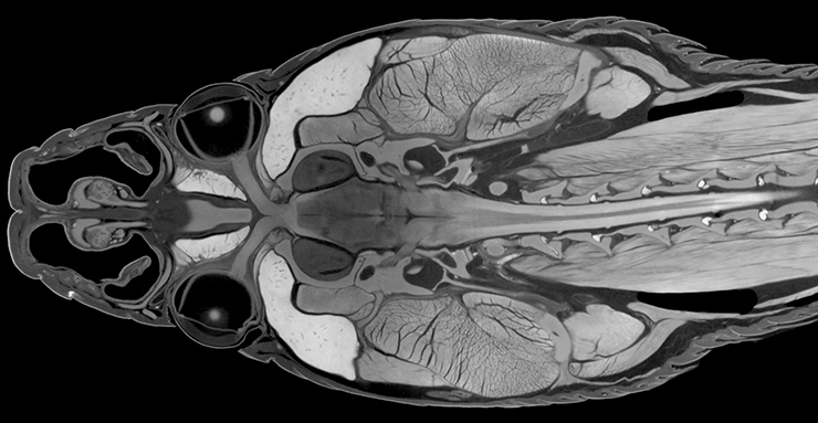
 Iodine-enhanced scan of a hatchling American alligator head, showing selected soft tissues. The light blue is the brain, purple shows cranial nerves, darker blue is the olfactory bulb, and orange shows eye muscles. Image by Paul Gignac and Nate Kley. |
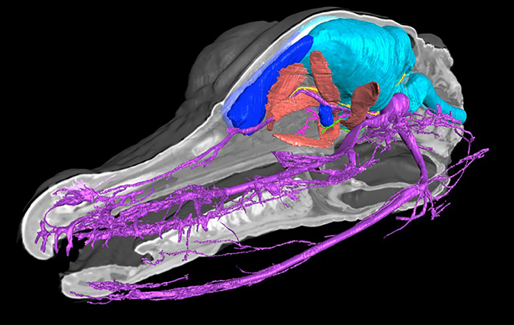
.jpg) Three-dimensional reconstruction of a juvenile giraffe head in lateral view, prepared using radiopaque vascular injection and CT imaging. This process differs slightly from diceCT in that the arterial system of this specimen was injected with barium-laced latex, which allows the vasculature to show up in considerable detail. For example, between the teeth in this image one can see the arteries of the tongue, part of which is sticking out of this giraffe’s mouth. This image was exhibited at the Tulsa LivingArts Gallery in 2017. Image by Haley O’Brien. |
.jpg) Color slide showing exterior and internal structure of a 3D-printed print head for a 3D printer. This new print head is based on a gear pump, which pumps fluid, and was designed for use in next-generation 3D printers that will allow for unlimited printing volume. Image by Haley O’Brien and Wenchao Zhou. |
|
|
.jpg)
 Cross-section of a rabbit jaw, focused on the temporomandibular joint, or “TMJ,” showing the internal structure of the bone and a color map representing bone thickness. Warmer colors (reds, oranges) show thicker bone while cooler colors (blues, purples) show thinner bone. This image was produced as part of Claire Terhune’s research on how diet is related to internal bone structure, and whether rabbits raised on different diets show differences in bone thickness. Image by Claire Terhune. |
 From Spiro, a Caddo archeological site in eastern Oklahoma, a woven “box” lid with copper plating (top) and 3D reconstruction of a cord-wrapped basket fragment with construction elements, including interlayering of plaited plant-fibers, split cane, and woven “yarn” or cordage made from plant fiber and animal fur. Inset shows close-up of braided cordage. Image by Haley O’Brien and George Sabo. |
|
|
.jpg)
To learn more about the consortium and system, and to view additional images, visit Research Frontiers.
Topics
Contacts
Claire Terhune, assistant professor
Department of Anthropology
479-856-3529, cterhune@uark.edu
Matt McGowan, science and research communications officer
University Relations
479-575-4246, dmcgowa@uark.edu